
This website uses cookies so that we can provide you with the best user experience possible. Cookie information is stored in your browser and performs functions such as recognising you when you return to our website and helping our team to understand which sections of the website you find most interesting and useful.
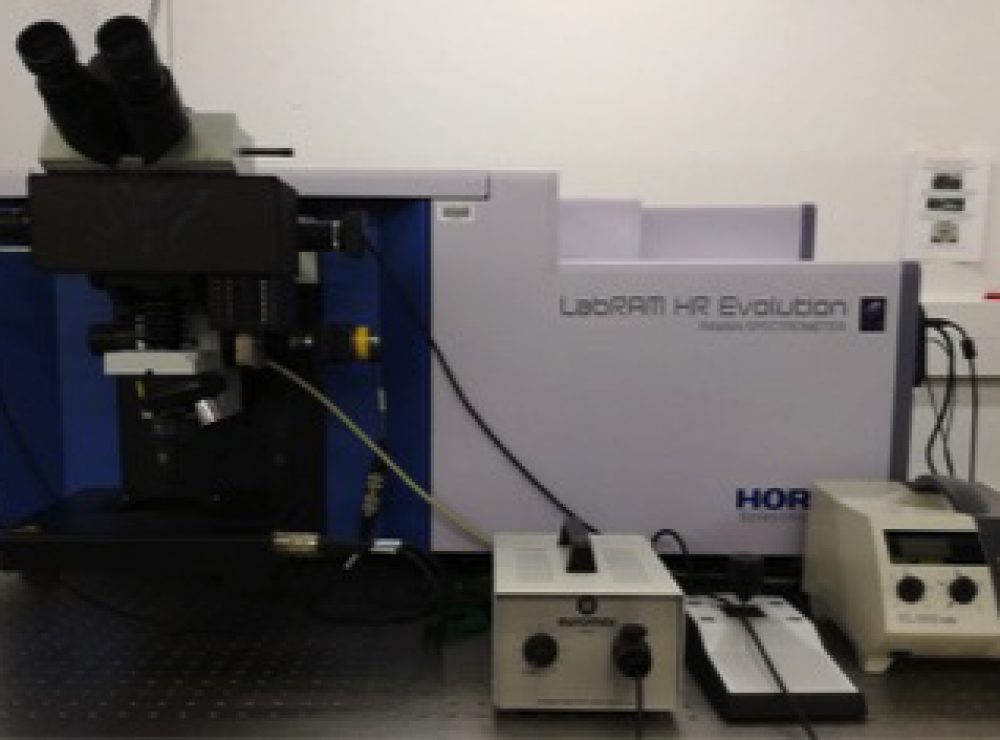
Fully integrated confocal Raman microscope instrument
- Partner:The University of Manchester
- Facility:Multidisciplinary Characterisation Facility (MCF)
Or call us now on 0161 275 8382
Detailed Description
Raman Spectroscopy can offer a powerful non-destructive and non-contact method to analyse the chemical composition and molecular structure of a material. The technique involves illuminating a sample with a monochromatic light source, such as a laser. Light that is scattered has a different colour to the light source and can be analysed using a highly sensitive spectrometer. Coupling of a confocal microscope to a Raman spectrometer enables surfaces to be mapped with high spatial resolution, giving an image of the uniformity (homogeneity) of the material.
Uses/Applications
The Horiba LabRAM Evolution HR Raman Spectrometer is a fully integrated confocal Raman microscope instrument with three laser sources (325nm, 488nm and 633nm), two diffraction gratings (600g/mm and 1800g/mm), motorised XYZ stage and free-space microscope. The instrument is controlled using LabSpec 6 software.
Laser Wavelength and Power: 325nm – 20mW, 488nm – 100mW (adjustable), 633nm – 17mW.
Diffraction Grating: 600g/mm and 1800g/mm
Laser (Rayleigh) Filtering Type: Edge
Microscope: Olympus BXFM Free-Space Microscope with trinocular head
Microscope Objectives: VISIBLE – 5X (NA 0.10), 10X (NA 0.25), 20X (NA 0.4), 50X SLWD (NA 0.35), and 100X (NA 0.90). UVB – 15X (NA 0.32) and 40X (NA 0.50).
Polarisation Options: Half-Wave Plate (VIS and NUV) for incident light and Analyzer (VIS and UV) for scattered light.
Spectral range: 220-1100nm (upgradable to 2200nm)
CCD: Open Electrode CCD air-cooled to -60°C
Märzhäuser mapping stage specification: Resolution XY = 100nm, Z = 10nm. Maximum range X = 75 mm – Y = 50 mm.
Duoscan mapping resolution: XY = 50nm
Rapid mapping mode: Swift ultra-fast mapping (max scan time per point = 1s)
Software: LabSpec 6